There’s More Than One Type Of Dental X-ray
May 5, 2020

X-rays are one of the most important tools in your dentist’s office. They allow care providers to get a better understanding of what’s actually happening inside your mouth, resulting in more accurate and effective treatment planning. But did you know that your dentist has more than one type of dental X-ray available to use? The type they choose depends on the information they are trying to collect. If you’re curious about the different kinds available, keep reading to find out more.
Bitewing
During your regular checkup exams, this is the type of X-ray you likely receive. A small wing shaped device containing a piece of film is placed inside your mouth to generate the image. With it, your dentist is able to see through the crowns of your teeth and the upper half of their roots in a portion of your mouth. They are helpful when your dentist is trying to determine the extent of the damage caused by a cavity.
Periapical
These are similar to a bitewing X-rays, except that they are capable of showing the entire length of tooth roots. You’ll know you’re getting one of these when your dentist has you bite down on a device that supports a large ring outside your mouth. Your dentist will use these X-rays if they suspect a cavity has damaged the interior tissues of a tooth, or if they want to evaluate the health of the bone around certain teeth.
Occlusal
This type of X-ray is used to view an entire arch of teeth at once. They are useful when your dentist is trying to track the movement of your teeth or when trying to find growths or injuries beneath the gum line. In order to take this type of X-ray, you will be asked to bite down directly on a sheet of film while the image is being generated.
Panoramic
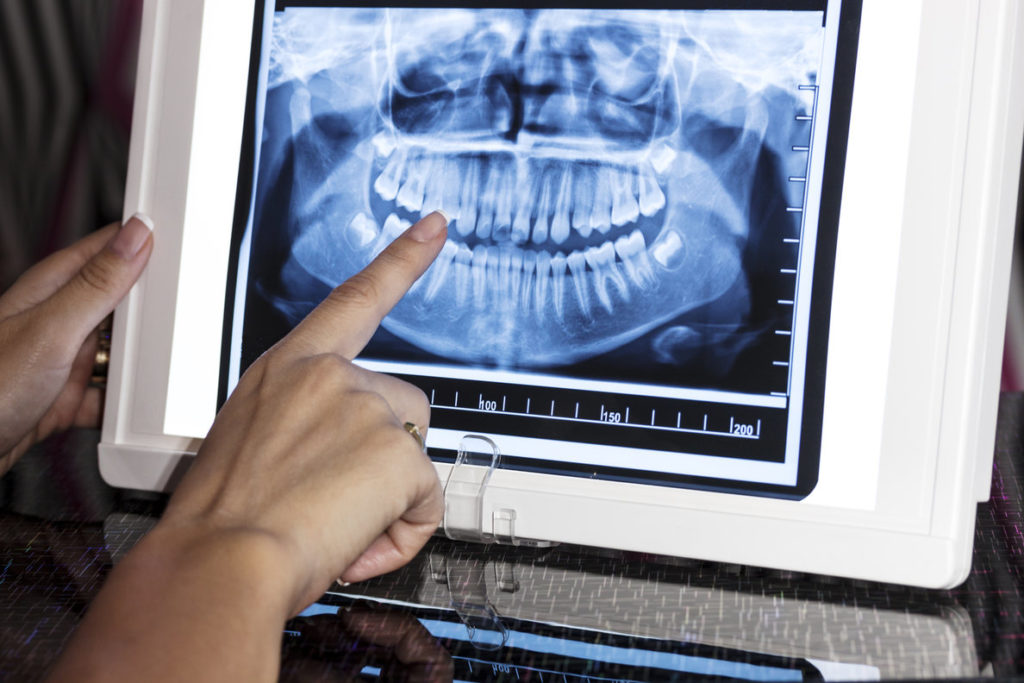
When your dentist wants to see all of the structures of your mouth at once, they will use a panoramic X-ray. In young adults, this type of X-ray can be helpful in planning for orthodontic care and wisdom teeth extractions. For older patients, it allows dentists to identify growths caused by oral cancer in areas that can’t be easily viewed during a dental exam.
To have one of these X-rays completed, your dentist will have you stand in a large specialized device. Once you bite down on a stabilizing plate in front of you, panels will rotate around your head to create a representation of your entire oral cavity.
Cone Beam CT Scan
A cone beam scan is the most advanced form of dental imaging technology in use today. The procedure to receive one is almost identical to the process used for a panoramic X-ray. However, the scan produces a three-dimensional image instead of a two-dimensional one. Your dentist will request this type of scan to identity problems in the bones around your mouth, as well as when planning the placement of dental implants.
Your dentist only wants to recommend treatment when it is absolutely necessary. Each type of X-ray allows them to better understand the specifics behind your dental issue and create a treatment plan that truly fits your needs.
About The Practice
Advanced Dental Care of Springfield believes in empowering patients so that they can be in control of their dental care. The team at the practice utilizes panoramic X-rays and cone beam CT scans so that patients can see what’s going on in their mouths. The unique combination of specialized expertise and modern technology at the practice allows the treatment team to handle nearly any dental issue in-house. If you’d like to start improving your oral health, you can make an appointment though the practice’s website or at 217-546-3333.
No Comments
No comments yet.
RSS feed for comments on this post.
Sorry, the comment form is closed at this time.
